Home » Without Label » Shoulder Muscles Diagram / Anatomy Chart Shoulder Girdle Muscles Diagram / The supraspinatus, the infraspinatus, the teres minor and the subscapularis.
Shoulder Muscles Diagram / Anatomy Chart Shoulder Girdle Muscles Diagram / The supraspinatus, the infraspinatus, the teres minor and the subscapularis.
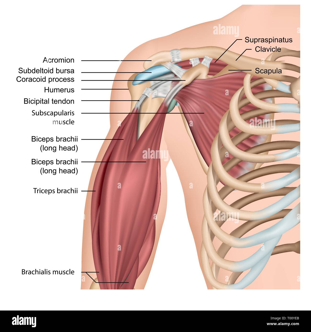
Shoulder Muscles Diagram / Anatomy Chart Shoulder Girdle Muscles Diagram / The supraspinatus, the infraspinatus, the teres minor and the subscapularis.. The shoulder joint is formed where the humerus (upper arm bone) fits into the scapula (shoulder blade), like a ball and socket. Shoulder muscles move the shoulder blades and upper arm bones. The back's muscles start at the top of the back (named the cervical vertebrae) and go to the tailbone (also named the coccyx). Insert on skin or another muscle action: The rotator cuff is a collection of muscles and tendons that surround the shoulder, giving it support and allowing a wide range of motion.
Diagram of the shoulder, including the location of the rotator cuff. Muscle anatomy back 12 photos of the muscle anatomy back back muscle anatomy images, back muscle anatomy of the human body, back pain muscle anatomy, muscle anatomy lower back, posterior back muscle anatomy, human muscles, back muscle anatomy images, back muscle anatomy of the human body, back pain muscle anatomy. The muscles of the shoulder support and produce the movements of the shoulder girdle.they attach the appendicular skeleton of the upper limb to the axial skeleton of the trunk. Muscles of the shoulder : As the name implies, the rotator cuff functions to allow you to rotate your shoulder and lift your arm.
Shoulder Muscles Anatomy High Resolution Stock Photography And Images Alamy from c8.alamy.com The main shoulder muscles are trapezius, deltoid, pectoralis major and 4 rotator cuff muscles: The back's muscles start at the top of the back (named the cervical vertebrae) and go to the tailbone (also named the coccyx). Back pain is one of the most common kinds of pain for adults, and muscle strains are the most common type of back pain. The muscles in the shoulder aid in a wide. These are located in the shoulder blade area, and each related tendon also attaches to the humerus. Four of them are found on the anterior aspect of the shoulder, whereas the rest are located on the shoulder's posterior aspect and in the back. See more ideas about muscle anatomy, shoulder muscle anatomy, shoulder muscles. The supraspinatus is located on the greater tubercle of the humerus.
As the name implies, the rotator cuff functions to allow you to rotate your shoulder and lift your arm.
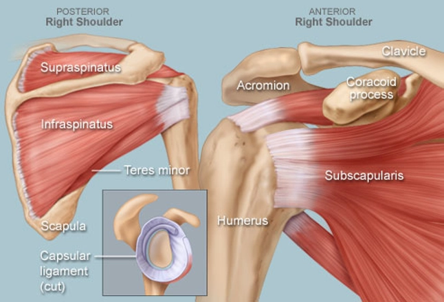
Located superior to the shoulder joint the deltoid muscle works with the supraspinatus to abduct the arm at the shoulder. On the anterior side of the shoulder the coracobrachialis serratus anterior pectoralis major and pectoralis minor muscles work as a group to flex and adduct the scapula and humerus anteriorly toward the sternum. Innervation of the latissimus is via the thoracodorsal nerve. The shoulder blade (scapula) connects to the collarbone (clavicle) at this joint. The bursa is a small sac of fluid that cushions and. The following is an overview of the shoulder muscle anatomy. What are common rotator cuff injuries? The rotator cuff is a group of four muscles and tendons that surround the glenohumeral joint. Contents hide deltoids anatomy. The rotator cuff is a group of four muscles and tendons that surround the glenohumeral joint. The supraspinatus is located on the greater tubercle of the humerus. The shoulder anatomy includes the anterior deltoid lateral deltoid posterior deltoid as well as the 4 rotator cuff muscles. Humurus is held on to scapular by tendons of muscles and it is the integrity of these muscles that keeps the shoulder joint intact (rotator cuff muscles) scapular=attachment for muscles of the back and shoulder and is fairly mobile parts:
The supraspinatus is located on the greater tubercle of the humerus. Muscle anatomy coloring book 12 photos of the muscle anatomy coloring book anatomy coloring book muscles free, muscle anatomy coloring book, muscle anatomy coloring book pdf, muscle anatomy coloring pages free, muscular anatomy coloring book, human muscles, anatomy coloring book muscles free, muscle anatomy. The muscles in the shoulder aid in a wide. Muscles of the shoulder : A muscle contracts to move bones;
Shoulder Human Anatomy Image Function Parts And More from img.webmd.com For that reason, and because of the dexterity of the shoulder joint itself, the musculature of the shoulder is complex, ranging from massive prime mover muscles to finer stabilizer and fixator muscles. The rotator cuff muscles are important stabilizers and movers of the shoulder joint. And the ligaments, which connect bones. The rotator cuff is a group of four muscles and tendons that surround the glenohumeral joint. The list of muscles and their functions are presented below. Numerous muscles help stabilize the three joints of. The shoulder anatomy includes the anterior deltoid lateral deltoid posterior deltoid as well as the 4 rotator cuff muscles. The shoulder joint is formed where the humerus (upper arm bone) fits into the scapula (shoulder blade), like a ball and socket.
Learn about these muscles, their origin and insertion points, and their functional anatomy.
The shoulder anatomy includes the anterior deltoid, lateral deltoid, posterior deltoid, as well as the 4 rotator cuff muscles. Related posts of diagram of shoulder muscles and tendons muscle anatomy back. The back's muscles start at the top of the back (named the cervical vertebrae) and go to the tailbone (also named the coccyx). The shoulder muscles consist of the deltoids and the rotator cuff group.the deltoids are the muscles that can be seen on the outside of the body, whilst the rotator cuff group is found within the shoulder joint itself, providing structural support and allowing the shoulder to perform many functions. The tendons, which anchor muscle to bone; Related posts of shoulder muscles and tendons diagram muscle anatomy coloring book. The rotator cuff is a collection of muscles and tendons that surround the shoulder, giving it support and allowing a wide range of motion. These are located in the shoulder blade area, and each related tendon also attaches to the humerus. The muscles of the shoulder support and produce the movements of the shoulder girdle.they attach the appendicular skeleton of the upper limb to the axial skeleton of the trunk. Numerous muscles help stabilize the three joints of. The shoulder is a complex combination of bones and joints where many muscles act to provide the widest range of motion of any part of the body. The main shoulder muscles are trapezius, deltoid, pectoralis major and 4 rotator cuff muscles: Insert on skin or another muscle action:
Related posts of shoulder muscles and tendons diagram muscle anatomy coloring book. Supraspinatus is the first shoulder muscle to initiate arm shoulder abduction. On the anterior side of the shoulder the coracobrachialis serratus anterior pectoralis major and pectoralis minor muscles work as a group to flex and adduct the scapula and humerus anteriorly toward the sternum. Parts of the right shoulder blade: Insert on skin or another muscle action:
Shoulder Anatomy Muscles Diagram Anatomy System Human Body Anatomy Diagram And Chart Images from anatomysystem.com The most common shoulder injuries are sprains, strains, and tears. Related posts of diagram of shoulder muscles and tendons muscle anatomy back. Muscle anatomy back 12 photos of the muscle anatomy back back muscle anatomy images, back muscle anatomy of the human body, back pain muscle anatomy, muscle anatomy lower back, posterior back muscle anatomy, human muscles, back muscle anatomy images, back muscle anatomy of the human body, back pain muscle anatomy. The shoulder muscles are associated with movements of the upper limb shoulder muscles diagram. Diagram of the shoulder, including the location of the rotator cuff. Parts of the right shoulder blade: Shoulder muscles move the shoulder blades and upper arm bones. These muscles form the outer shape of the shoulder and underarm.
The rotator cuff is a group of four muscles and tendons that surround the glenohumeral joint.
The muscles of the shoulder are associated with movements at the shoulder joint. Back pain is one of the most common kinds of pain for adults, and muscle strains are the most common type of back pain. Shoulder flexion is movement of the shoulder in a forward motion. Shoulder mri radiographical and illustrated anatomical atlas from www.imaios.com While seated, have your partner place one hand at the front of your shoulder joint and one hand at the rear. The bursa is a small sac of fluid that cushions and. The back's muscles start at the top of the back (named the cervical vertebrae) and go to the tailbone (also named the coccyx). The shoulder joint is formed where the humerus (upper arm bone) fits into the scapula (shoulder blade), like a ball and socket. Shoulder muscles move the shoulder blades and upper arm bones. The shoulder muscles and shoulder tendons involved with shoulder mobility include the four rotator cuff muscle and tendon pairs: Learn about these muscles, their origin and insertion points, and their functional anatomy. It is the most common cause that causes pain between the shoulder blades. See more ideas about muscle anatomy, shoulder muscle anatomy, shoulder muscles.